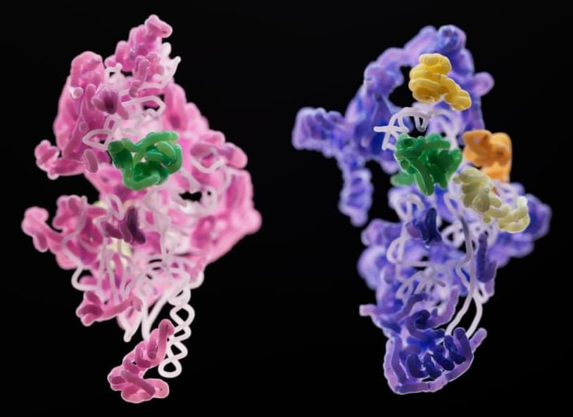
The findings shed a rare light on mitoribosomes, the unique ribosomes found within the cell’s mitochondria. Ribosomes, the tiny protein-producing factories within cells, are ubiquitous and look largely identical across the tree of life. Those that keep bacteria chugging along are, structurally, not much different from the ribosomes churning out proteins in our own human cells.
But even two organisms with similar ribosomes may display significant structural differences in the RNA and protein components of their mitoribosomes. Specialized ribosomes within the mitochondria (the energy producing entities within our cells), mitoribosomes help the mitochondria produce proteins that manufacture ATP, the energy currency of the cell.
Scientists in the laboratory of Sebastian Klinge wondered how mitoribosomes evolved, how they assemble within the cell, and why their structures are so much less uniform across species. To answer these questions, they used cryo-electron microscopy to generate 3D snapshots of the small subunits of yeast and human mitoribosomes as they were being assembled. Their findings, published in Nature, shed light on the fundamentals of mitoribosome assembly, and may have implications for rare diseases linked to malfunctioning mitoribosomes.


Leave a reply